
What Happens During a Routine Eye Exam?
Posted by: Pepose Vision Institute in Eye Care on February 6, 2026
Your eyes do more than help you see clearly. They can reveal early signs of serious health conditions like diabetes, high blood pressure, glaucoma, and macular degeneration.
Regular eye exams catch these problems before they affect your vision or overall health. Keep reading to learn exactly what happens during a comprehensive eye exam and why each step protects your long-term vision health.
Why Are Routine Eye Exams Important?
During a routine eye exam, your eye doctor examines the health of your entire visual system, looking for early warning signs of eye diseases that often have no symptoms in their early stages. Conditions like glaucoma, cataracts, and age-related macular degeneration can progress silently for years before you notice vision changes. Finding them early means more treatment options and better outcomes.
Beyond eye-specific conditions, your eyes offer a unique window into your overall health. During a comprehensive eye exam, your ophthalmologist can spot signs of diabetes, high blood pressure, autoimmune diseases, and even some types of cancer. Blood vessels in your retina are the only blood vessels in your body that doctors can view directly without surgery. This makes routine eye exams an important part of your preventive healthcare.
Adults with healthy vision typically need comprehensive exams every one to two years, while those with existing eye conditions, diabetes, or family history of eye disease may need annual visits. Children should have their first exam around age three and then before starting school.
At Pepose Vision Institute, the team creates personalized exam schedules based on your individual risk factors and vision needs.
What to Expect When You Arrive
Your routine eye exam begins before you even meet with the doctor.
When you check in, you’ll complete a patient history form or update your existing information. This paperwork asks about current vision problems you’re experiencing, like blurry vision, eye strain, or difficulty seeing at night. You’ll also list any medications you take and note any eye injuries or surgeries you’ve had in the past.
Your eye care team will ask about your family’s eye health history, too. Many eye conditions run in families, so knowing if your parents or siblings have glaucoma, macular degeneration, or other eye diseases helps your doctor assess your risk.
This conversation also covers your general health, including conditions like diabetes or high blood pressure that can affect your eyes.
Tests for Vision and Refraction
Visual Acuity Test
The exam itself starts with the familiar eye chart test.
You’ll cover one eye at a time and read letters that get progressively smaller on a chart across the room. This visual acuity test measures how clearly you see at a distance. The results are recorded as a fraction, like 20/20 or 20/40.
Here’s what those numbers mean: if you have 20/40 vision, you see at 20 feet what a person with perfect vision sees at 40 feet. In other words, objects need to be closer for you to see them clearly.
Your eye doctor tests each eye separately because your two eyes may have different vision strengths.
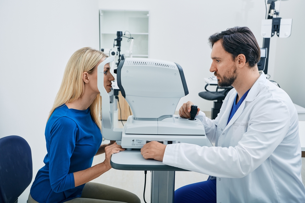
Refraction Assessment
Next comes the refraction test, which determines your exact prescription for glasses or contact lenses. You’ll look through a device called a phoropter while your doctor flips between different lens options. You’ve probably heard the classic question: “Which is better, one or two?” Your answers help your eye doctor fine-tune your prescription.
The phoropter contains hundreds of lens combinations that test for nearsightedness, farsightedness, and astigmatism. This process takes several minutes as your doctor narrows down the perfect lens strength for each eye.
Some offices now use computerized refraction that automatically measures your prescription, though most doctors still verify these results manually for accuracy.
Tests for Evaluating Your Eye Health
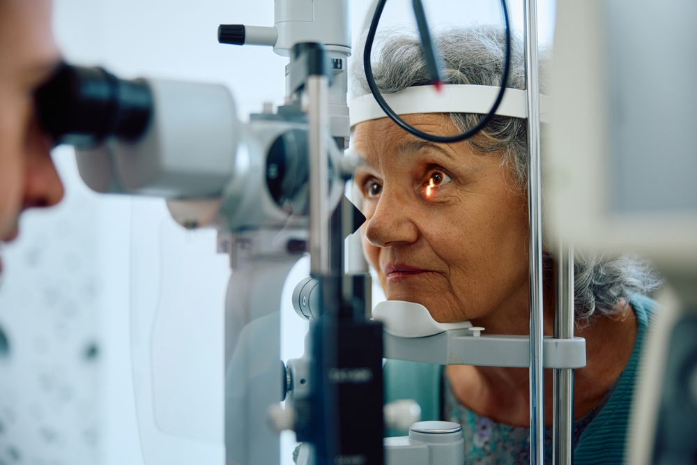
Pupil Dilation
For a complete view of your eye health, your doctor needs to examine the inside of your eyes. This requires dilating your pupils with special eye drops. The drops take about 15 to 30 minutes to work fully. You might feel a slight sting when the drops go in, but it passes quickly.
Dilated pupils let your ophthalmologist see the back of your eye, including your retina, optic nerve, and blood vessels. Yes, bright lights will bother your eyes more than usual while you’re dilated, and you might have some trouble focusing on nearby objects.
The effects typically wear off within four to six hours, though some people remain sensitive to light for the rest of the day.
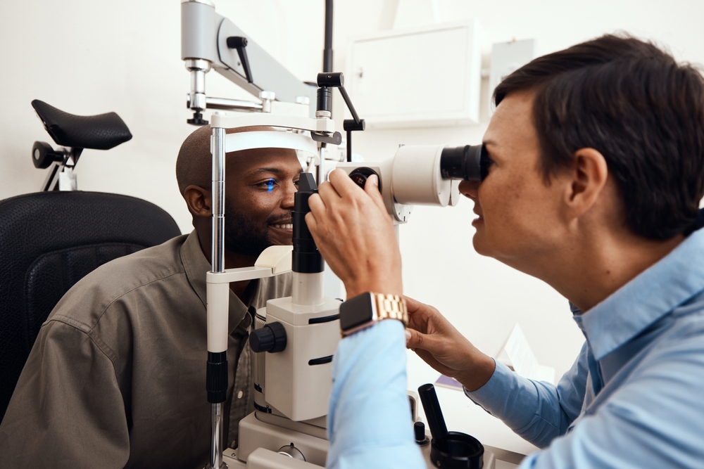
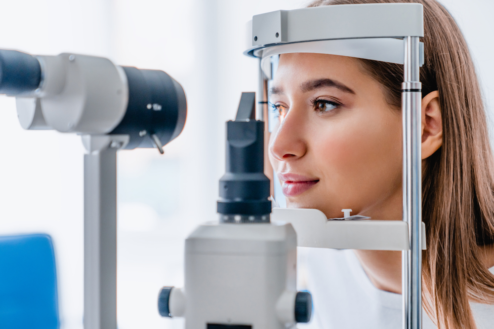
Retinal Examination
Once your pupils are dilated, your doctor examines your retina using a special magnifying lens and bright light.
The retina is the light-sensitive tissue at the back of your eye that sends visual signals to your brain. During this examination, your ophthalmologist looks for signs of retinal tears, detachment, macular degeneration, and diabetic retinopathy.
Your doctor also checks your optic nerve, which connects your eye to your brain. The optic nerve’s appearance can reveal signs of glaucoma, optic neuritis, or increased pressure around your brain.
Blood vessels throughout your retina are examined for narrowing, bleeding, or swelling that might indicate high blood pressure, diabetes, or other systemic conditions. This part of the exam takes just a few minutes but provides information about both your eye health and overall wellness.
Tonometry (Eye Pressure Test)
Measuring the pressure inside your eyes is a key part of glaucoma screening. High eye pressure can damage your optic nerve over time, leading to vision loss. The tonometry test measures this internal pressure quickly and painlessly.
You might have experienced the “air puff” test, where a machine blows a small puff of air at your eye while you look at a light. Some people find this test startling, but it doesn’t hurt.
Other methods involve gently touching the surface of your numbed eye with a small probe. Your doctor chooses the method that works best for getting accurate readings. Normal eye pressure ranges from 10 to 21 mmHg, though what’s normal varies from person to person.
Additional Specialized Tests
Visual Field Testing
Your peripheral vision (side vision) is just as important as your central vision.
A visual field test maps your complete field of vision to detect blind spots you might not have noticed. You’ll look at a screen or inside a bowl-shaped instrument while lights flash in different locations. You press a button each time you see a light.
This test helps detect glaucoma, strokes, brain tumors, or other neurological conditions that affect vision. The computerized test takes about 10 minutes per eye. Staying focused and responding quickly gives your doctor the most accurate results.
Optical Coherence Tomography (OCT)
Many eye care offices now use OCT imaging as part of comprehensive exams. This technology works like an ultrasound for your eyes, using light waves instead of sound waves to create detailed cross-sectional images of your retina. OCT scans can detect subtle changes in your retinal layers that aren’t visible during a regular examination.
The test itself is simple and fast. You rest your chin in a support and look at a target light while the machine scans your eye.
The scan takes just seconds and doesn’t require dilation or touching your eye. OCT imaging is particularly valuable for monitoring macular degeneration, diabetic retinopathy, and glaucoma progression.
Ready to schedule your routine eye exam? Contact Pepose Vision Institute in Chesterfield, Missouri, today for expert, compassionate eye care that keeps your vision clear and healthy for years to come.