Corneal Transplants St Louis | Cornea Replacement St Louis, MO

Corneal Transplants

Illustration of an Eye Showing the Cornea

The cornea is the clear tissue covering the front of your eye. Like the crystal on your watch, your cornea serves as a protective window that allows light to enter your eye and come to focus on your retina. The cornea must remain clear for you to have good vision.

Corneal disease, degeneration or scarring from injury can cause the cornea to become cloudy and blur your vision. If corneal problems make it hard for you to see well enough to perform normal activities, such as reading or driving, a corneal transplant might help restore your vision.

A cornea transplant is a surgical procedure to replace part of your cornea with corneal tissue from a donor. A cornea transplant can restore vision, 

reduce pain and improve the appearance of a damaged or diseased cornea. Also called keratoplasty, is typically performed as an outpatient procedure.

The doctors at Pepose Vision Institute are leading cornea specialists and have an excellent track record in corneal transplants. This translates into:

  • Faster visual recovery: little to no discomfort
  • Less need for repeat surgery to achieve best vision result possible
  • Minimized risk of complications and transplant rejection that can compromise your sight
  • The greatest amount of functional vision possible in the treated eye

When is a corneal transplant needed?

A corneal transplant is typically needed when:

  • Vision cannot be corrected satisfactorily using other medical approaches
  • Painful swelling cannot be relieved by medications or special contact lenses
  • Corneal failure occurs after other eye surgery, such as cataract surgery
  • Severe Keratoconus, a steep curving of the cornea, develops
  • Hereditary corneal failure, such as Fuchs’ dystrophy, exists
  • Scarring after infections, especially after herpes, occurs
  • Rejection occurs after first corneal transplant
  • Scarring arises after injury

Only an experienced, cornea subspecialty trained surgeon can appropriately determine if and when a transplant will enhance the quality of your vision.


We always provide our patients with compassionate and customized care.


How is a corneal transplant performed?

A cornea transplant is performed under local or general anesthesia at an outpatient surgery center. When we use local anesthesia, you may be able to hear what is going on, even though your eye and face will be numb and you may be groggy. Our cornea subspecialty trained surgeon will remove the hazy part of your cornea and a clear cornea from a donor will be put in its place. Tiny stitches, about 1/3 the diameter of a human hair, are used to hold this tissue in place. The procedure usually lasts for to two hours, depending on whether any additional procedures are done to remedy other vision problems in the eye.


Frequently asked questions

The cornea is the clear front of the eye that covers the colored iris and the round pupil. Light is focused onto the retina at the back of the eye by passing through the cornea. If the cornea is not clear or shaped properly, then this light will not be focused properly and vision problems will arise. In the extreme case, a badly damaged cornea can lead to blindness.

Your cornea may become hazy or damaged because of disease or injury. Corneal infections caused by inadequately cleaned contact lenses can lead to permanent corneal damage if not treated promptly. Trauma to the eye can likewise permanently injure the cornea, as can certain progressive diseases, such as keratoconus. When the cornea is damaged, it may become swollen or scarred, and its smoothness and clarity may be lost. The scars, swelling or resulting irregular shape cause the cornea to scatter or distort light, resulting in glare or blurred vision. Like a foggy window, light no longer passes through it well, so a blurred image forms in your eye.

Corneal transplants have been performed in this country for over 50 years. They account for the single largest number of transplant procedures in the U.S. each year. There have been exciting innovations in the way in which such transplants are performed, thereby increasing the success rate and reducing tissue rejection by recipients. Pepose Vision Institute’s cornea subspecialty trained surgeons have contributed a great deal to the development of these new technologies, conducting clinical studies in order to bring the latest advances to their cornea patients.

We have had babies as young as 3 months old who have required corneal transplants because of congenital cornea defects. Through cornea transplants, we have saved the sight of young athletes who have suffered trauma to their corneas. We have performed corneal transplants on patients of all ages who have seriously impaired corneas.

A corneal transplant is typically needed when:

  • Vision cannot be corrected satisfactorily using other medical approaches
  • Painful swelling cannot be relieved by medications or special contact lenses
  • Corneal failure occurs after other eye surgery, such as cataract surgery
  • Severe Keratoconus, a steep curving of the cornea, develops
  • Hereditary corneal failure, such as Fuchs’ dystrophy, exists
  • Scarring after infections, especially after herpes, occurs
  • Rejection occurs after first corneal transplant
  • Scarring arises after injury

Only an experienced, cornea subspecialty trained surgeon can appropriately determine if and when a transplant will enhance the quality of your vision.

A cornea transplant is performed under local or general anesthesia at an outpatient surgery center. When we use local anesthesia, you may be able to hear what is going on, even though your eye and face will be numb and you may be groggy. Our cornea subspecialty trained surgeon will remove the hazy part of your cornea and a clear cornea from a donor will be put in its place. Tiny stitches, about 1/3 the diameter of a human hair, are used to hold this tissue in place. The procedure usually lasts for to two hours, depending on whether any additional procedures are done to remedy other vision problems in the eye.

The operation is virtually painless. Anesthesia is either local or general, depending on your age, medical condition and eye disease. You will not see the surgery while it is happening, and will not have to worry about keeping your eye open or closed.

No. The cornea is a clear tissue that is in the front of the eye and the iris, which gives your eyes their color.

Cornea tissue is subjected to a battery of laboratory tests prior to its release for transplant purposes. Pepose Vision Institute’s Medical Director, Dr. Jay Pepose, is the Medical Director of MidAmerica Tissue Services, and has had decades of experience in validating the safety of corneal tissue. As an M.D. and Ph.D. with specialization in microbiology and infectious diseases, Dr. Pepose has conducted landmark medical research that has demonstrated that corneal tissue does not transmit the AIDS virus or many other viruses that are of concern in the organ transplant community. In addition to the numerous tests that are performed on donor corneal tissue, Dr. Pepose reviews a variety of donor characteristics before accepting tissue for a transplant.

After your corneal transplant, you will be given eye medications that will prevent infection and reduce the chances of tissue rejection, when used as directed. Usually, matching donor and recipient by age, gender, race, or any other characteristic does not reduce the possibility of rejection. Donor tissue is rejected 5% to 30% of the time. The rejected cornea clouds and vision deteriorates. Most rejections, if treated promptly, can be stopped with minimal injury. Warning signs of rejection are: persistent discomfort; light sensitivity; redness; or change in vision. In the case where rejection does occur, a corneal transplant can be repeated, usually with good results. In some instances, matching the blood types of donor and recipient helps when there has been previous corneal tissue rejection. Still, the overall rejection rate for repeated transplants is higher than for the first time around, probably because of an underlying factor that causes a patient to be more prone to rejection.


Caitlin with Dr. Pepose

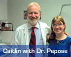
Corneal Transplant Case Study: Little Caitlin Kuehling was born with a condition called Peter’s Anomaly – in which the corneas are cloudy and the iris is misshapen. Essentially, she was functionally blind. Dr. Pepose knew that of the 40,000 cornea transplant operations in the country at the time, only 300 had been performed on babies. It was a risky procedure for an eye so small. But Caitlin’s mother Monica just wanted for her baby what other children had: “To see the moon and the stars and more.” It’s been more than 16 years since her operation, and that’s just what “Baby” Caitlin can now see. The moon. The stars. And all the beauty the world has to offer.


Why choose Pepose Vision for your corneal transplant surgery?

The eye doctors of Pepose Vision Institute have one of the best track records of protecting and restoring our patients’ functional vision of any cornea specialist in the country.

This translates into:

  • Faster visual recovery: little to no discomfort.
  • Less need for repeat surgery to achieve best vision result possible.
  • Minimized risk of complications and transplant rejection that can compromise your sight of functional vision possible in their treated eye.

Experience

Cornea surgery outcomes improve dramatically if the surgeon is cornea subspecialty trained. It’s important to know that only cornea subspecialists at Pepose Vision Institute will perform your cornea procedure. Dr. Jay Pepose was recruited to Washington University/Barnes Hospital almost two decades ago because of his skill, training and experience in treating cornea-related injuries and disease.

Testing

Each cornea patient undergoes an intensive state-of-the-art testing process, directly supervised by our cornea subspecialty trained surgeons who then create a precise, personalized treatment plan.

Technology

Pepose Vision is always at the forefront of ophthalmic technology. As a research center, we often have access to technologies before most anyone else. We are leaders, both locally and nationally, in corneal tissue procurement and evaluation, relying on our state-of-the-art technologies to make sure that the donor tissue is safe and reduce risk of rejection.

Lifetime Commitment

Our surgeons vigilantly monitor the status of your eyes for the rest of your life to ensure that your vision continues to be its best, years AFTER your cornea surgery.

We recommend that the best way to ensure a successful outcome to your corneal transplant is to choose a highly experienced surgeon who is involved in developing state-of-the-art treatments to enhance your vision outcome.


Corneal Technology Leadership

Dr. Jay Pepose is one of the foremost cornea-subspecialty trained surgeons in the region and across the country. He has done pioneering work on tissue rejection and has served as Medical Director of MidAmerica Tissuebank Services, located in St. Louis. Dr. Pepose has performed thousands of corneal procedures, restoring and preserving the vision of infants, athletes and patients from all walks of life. The key to his success, and that of his team of cornea-trained surgeons, has been an uncanny ability to use the results of sophisticated diagnostic technologies to determine the appropriate surgical treatment for each cornea patient. Moreover, our cornea-trained surgeons are on-call 24/7, 365 days a year, so that any concerns can be addressed before they become major complications that threaten the initial transplant.

Because outstanding vision outcomes depend upon high-quality, precise preoperative analysis of your eyes, as well as advanced treatments and surgical techniques, we encourage you to learn more about:

Our State-Of-The-Art Diagnostic Tests

Ultrasonic Pachymetry produces reliable and reproducible measurements of corneal thickness. While Pepose Vision Institute begins its eye examination with this standard test, it progresses rapidly to the latest state-of-the-art technologies that provide additional information and confirmation of your corneal damage or disease.

Orbscan

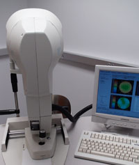
PVI is one of the few centers in the nation to evaluate all of its patients using both the Orbscan and Atlas corneal topography instruments. Topography is a way to measure the shape of the cornea. We evaluate your cornea at over 8,000 points and the various elevations are then represented color maps. Just as the height of land can be viewed from a helicopter circling above, the elevated peaks of the cornea can be printed in hot colors such as red and orange, and the valleys printed out in cool colors such as blue.

We measure both the front and back surfaces of the cornea. Its thickness at all locations can be plotted out, and a 3 dimensional reconstruction of the shape of the eye can be created that can be rotated and viewed by our surgeons at all angles. The Atlas topography device provides specialized computerized screening to evaluate the progression of a cornea condition called keratoconus.

Both diagnostic instruments give an extremely detailed depiction of the state of your cornea, including surface defects, scarring and thinning.

Historically, eye doctors measured the eye’s length by applying a probe to compress the front of the eye. With A-Scan Biometry, we use a gentle water bath technique called Immersion A-Scan. With this newest technology, the ultrasound probe does not come into direct contact with the cornea. We use this diagnostic test to confirm your intraocular lens calculations as well as assess lens thickness. It is especially useful in cases where patients have very dense cataracts.

Our Advanced Treatement and Procedures

Corneal transplants entail removal of your diseased or damaged tissue, and replacement with the donor tissue. After taking measurements of the amount of tissue to be removed, the diseased cornea is safely cut and lifted away from the eye. The donor tissue is cut to a matching size and placed on the patient’s eye. It is held in place with very fine stitches that may be removed anytime from one month to a year after surgery. If there is also a cataract present, this can be removed at the same time and replaced with an intraocular lens. At the completion of your transplant surgery, a patch and a metal shield are placed over your eye to protect it.

Pepose Vision Institute performs most of its corneal transplants using local anesthesia, thereby enhancing the speed of recovery and avoiding complications inherent in the use of general anesthesia. As tissue banks have grown and distribution networks become more sophisticated, corneal transplants can now be “scheduled” most of the time. Only rarely must a transplant be delayed or rescheduled due to the lack of acceptable tissue. We perform most of our transplants at an outpatient surgery center, depending upon a patient’s health status and surgical risk. The operation usually lasts between 1 and 2 hours, and most patients are discharged after a short stay in the recovery room. Your post-operative care will be intensive: our surgeons partner with you to ensure that very best vision outcome from your transplant. As soon as your vision stabilizes, we will fit you with glasses or contact lenses to maximize your vision potential.

Eye drops and ointment will be prescribed to avoid infection and minimize the risk of tissue rejection. The eye drops are required for at least a year and more often longer, to prevent rejection of the transplanted cornea. It is imperative that you comply with your medication schedule to assure the very best outcome from your corneal transplant.

DSAEK is a surgical technique for corneal transplantation. It permits our surgeons to transplant only the back layer of the cornea when medically indicated. This procedure may lead to a faster visual recovery than the traditional “full thickness” transplant. It requires fewer sutures and may induce less astigmatism. In the DSAEK procedure, a thin piece of donor corneal tissue containing endothelial cells and posterior stroma is placed in the eye through a small incision on the side of the eye. This tissue adheres to the back surface of the old cornea and clears the corneal swelling. Since we use a smaller incision than in “full thickness” transplants, the eye is left stronger so that if it is inadvertently struck there is less likelihood of a serious injury.

In order to make the new tissue stick onto the back of the old cornea, an air bubble is used to completely fill the front part of the eye for an hour after surgery. After that time the air is partially released and left overnight. During the first twenty-four hours, patients must stay on their back as much as possible to keep the air bubble positioned correctly, anchoring the new tissue in place.

Like full thickness corneal transplantation, eye drops are required for at least a year and more often for the rest of your life to prevent graft rejection. Similarly, you will need to wear eyeglasses or contact lenses after the transplant, in order to maximize your vision potential.

DMEK is the newest technique in corneal transplantation to replace the inner layer or ‘endothelium’ of the cornea. As with DSAEK, this procedure is most commonly performed for patients with Fuchs dystrophy, but other indications include corneal edema or swelling secondary to complicated cataract surgery, glaucoma surgeries and some intraocular lenses. DMEK involves a much thinner transplant than with DSAEK and with less tissue, the rejection rate is lower. This allows patients to safely switch to a lesser potent steroid. This is very important in patients who have glaucoma and whose eye pressure can be elevated by a steroid.

The recovery period is quicker than DSAEK and certainly quicker than PKP or “full thickness” cornea transplants. Once the diseased inner layer is removed, the donor transplant is injected into the eye and unfolded. Like DSAEK, air or gas is placed in the eye in order to ‘push’ the transplant against the back of the cornea. It is important to maintain face up positioning for the first three to five days to allow for the transplant to stick to the back of the cornea. Visual recovery is more similar to cataract surgery than to traditional corneal transplants, with vision usually improving within 2-4 weeks. Vision, both objective and subjective, is consistently better with DMEK.

Dr. Jay Pepose was the first in the entire nation to perform an amniotic membrane transplant. This type of surgical intervention may be performed in situations in which spontaneous healing of the surface of the cornea fails to occur, such as in cases of chemical burns, or where the cornea is severely infected. It is not the procedure of choice when the cornea is severely damaged, such as when a perforation occurs. The amniotic membrane, which surrounds the fetus in the womb, is a unique source of ‘biological dressing’ that has been used in wound healing for decades. These membranes appear to contain large amounts of growth factors (which encourage tissue regeneration), as well as anti-bacterial and anti-scarring properties. They are especially useful as tissue grafts because they do not encourage autoimmune rejection (as happens in organ transplant, for example). Amniotic membrane transplants have also been successful in promoting the growth of corneal tissue and in preventing corneal scarring. Within two months, the amniotic membrane tissue “dissolves,” leaving healed corneal tissue in its stead.

The amniotic membrane is “harvested” at the time that a baby is born, when it is no longer needed to sustain the baby. In this way, the miracle of birth can provide the miracle of sight to our cornea patients.


What’s your next step?

If you have any vision issues, please come see us.
Our experienced doctors will use advanced diagnostics to determine exactly how to help you see your very best, patiently answer your questions and carefully explain all of your options.

Arrow Pointing Up